
Keratoconus
Learn how keratoconus changes the shape of the cornea, which symptoms to watch for, and how Modern Eye Care helps patients manage vision changes with modern diagnostics and specialty lens options.
What Is Keratoconus ?
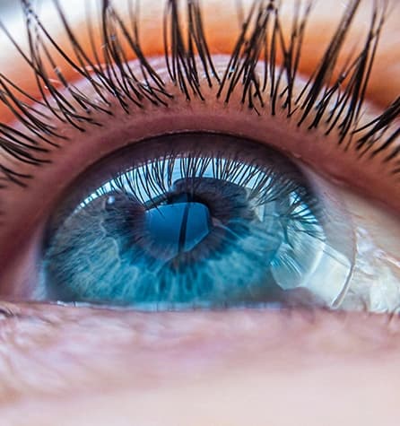
Keratoconus is a degenerative eye disease that causes the cornea to thin and bulge into an irregular cone-like shape. Because the cornea helps focus light entering the eye, these changes can lead to significant visual distortion. Keratoconus most often appears during the late teenage years, although it can also be diagnosed later in adulthood. It has been estimated to affect roughly one out of every 2,000 people, and it can occur across all geographic and demographic groups.
What Are The Risk Factors For Keratoconus?
Genetics. Patients with a family history of keratoconus or with certain systemic disorders, such as Down syndrome, are at a higher risk of developing keratoconus.
Chronic eye inflammation. Constant inflammation from allergies or irritants can contribute to the destruction of corneal tissue that may result in developing keratoconus.
Eye rubbing. Chronic eye rubbing is associated with developing keratoconus. It may also be a risk factor for disease progression.
Age. Keratoconus is often discovered in the teenage years. Generally, young patients with advanced keratoconus are more likely to need some form of surgical intervention as the disease progresses.
What Are The Symptoms Of Keratoconus?
Many keratoconus patients are unaware they have the disease, but it can often be diagnosed with modern eye exam technology. One of the earliest signs is slight blurring of vision or vision that becomes progressively harder to correct.
Other symptoms of keratoconus include:
- Glare and halos around lights
- Difficulty seeing at night
- Eye irritation or headaches associated with eye pain
- Increased sensitivity to bright light
- Sudden worsening or clouding of vision
How Is Keratoconus Diagnosed?
In addition to a complete medical history and eye exam, Dr. Sabahi performs the following tests to diagnose keratoconus:
- Corneal topography. This is the most accurate way to diagnose early keratoconus and follow its progression. A computerized image is taken that creates a map of the curve of the cornea.
- Slit-lamp exam. This examination of the cornea can help detect abnormalities in the outer and middle layers of the cornea.
- Pachymetry. This test is used to measure the thinnest areas of the cornea.
How Is Keratoconus Treated?
Treatment of keratoconus focuses on correction of vision and depends on the stage of the disease.
Dr. Sabahi often treats her patients with Scleral lenses. Scleral contact lenses are prescribed for a variety of irregular cornea conditions. The lens was designed using the latest research on the shape of the eye.They are designed to vault or rise over the irregular cornea condition and land on the white part of the eye (called the sclera). The scleral lens is made from a gas permeable material. They are available in single vision and multifocal lenses.
Available In Single Vision And Multifocal Lenses
This overview highlights different contact lens options.
SynergEyes VS™ Is Available In Single Vision And Multifocal Lenses
Progressive keratoconus can be treated by corneal collagen cross-linking. This one-time, in-office procedure involves the application of a vitamin B solution to the eye, which is then activated by ultraviolet light for about 30 minutes or less. The solution causes new collagen bonds to form, recovering and preserving some of the cornea's strength and shape.While the treatment cannot make the cornea entirely normal again, it can keep vision from getting worse.
Cross-linking was approved as a treatment for keratoconus by the FDA in April 2016, after clinical trials showed that it stopped or produced a mild reversal in bulging of the cornea within three to 12 months after the procedure.
Can Keratoconus Be Prevented?
There is no known prevention for keratoconus.